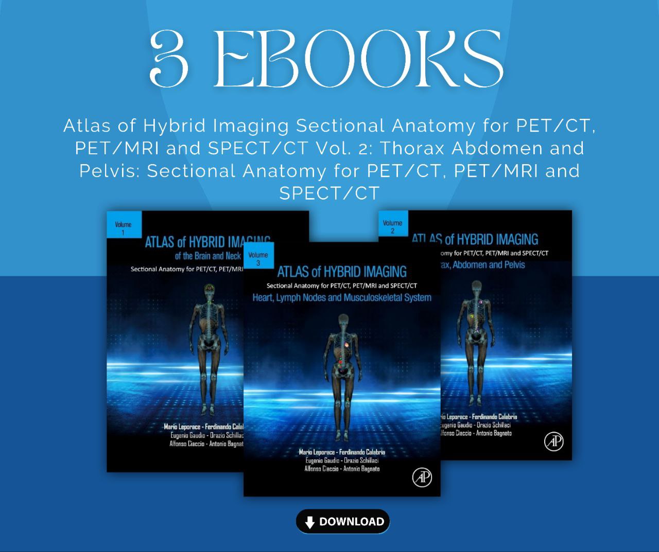
*The Hybrid Imaging Sectional Anatomy Atlas* is a three-volume collection designed to provide precise information on sectional anatomy for modern imaging techniques such as PET/CT, PET/MRI, and SPECT/CT. These books are indispensable references for professionals in the medical and imaging fields.
1. *Atlas of Hybrid Imaging Sectional Anatomy for PET/CT, PET/MRI, and SPECT/CT Vol. 1: Brain and Neck*
– *Overview*: This volume focuses on the anatomy of the brain and neck, offering detailed illustrations and high-quality images.
– *Contents*: Covers essential anatomical structures, aiding physicians in accurately interpreting scans and enhancing a comprehensive understanding of organ functions.
2. *Atlas of Hybrid Imaging Sectional Anatomy for PET/CT, PET/MRI, and SPECT/CT Vol. 2: Thorax, Abdomen, and Pelvis*
– *Overview*: This volume addresses the anatomy of the thorax, abdomen, and pelvis, emphasizing how to apply this knowledge in hybrid imaging.
– *Contents*: Includes over 1,000 illustrations and color images, providing crucial information about diseases and clinical procedures.
3. *Atlas of Hybrid Imaging Sectional Anatomy for PET/CT, PET/MRI, and SPECT/CT Vol. 3: Heart, Lymph Nodes, and Musculoskeletal System*
– *Overview*: This volume focuses on the anatomy of the heart, lymph nodes, and musculoskeletal system, facilitating an understanding of the complex interactions between these systems.
– *Contents*: Features updated diagnostic algorithms and treatment tables, making it an excellent reference for physicians and practitioners.
### Why Choose This Set?
– *Reliable Sources*: Written by experts in the field of medicine and imaging, ensuring accurate and trustworthy information.
– *User-Friendly Design*: Well-organized content allows for quick access to necessary information.
– *Educational Tools*: Ideal for students and practitioners looking to expand their knowledge in medical imaging.
*Get this collection today and elevate your anatomical knowledge!*


Reviews
There are no reviews yet.