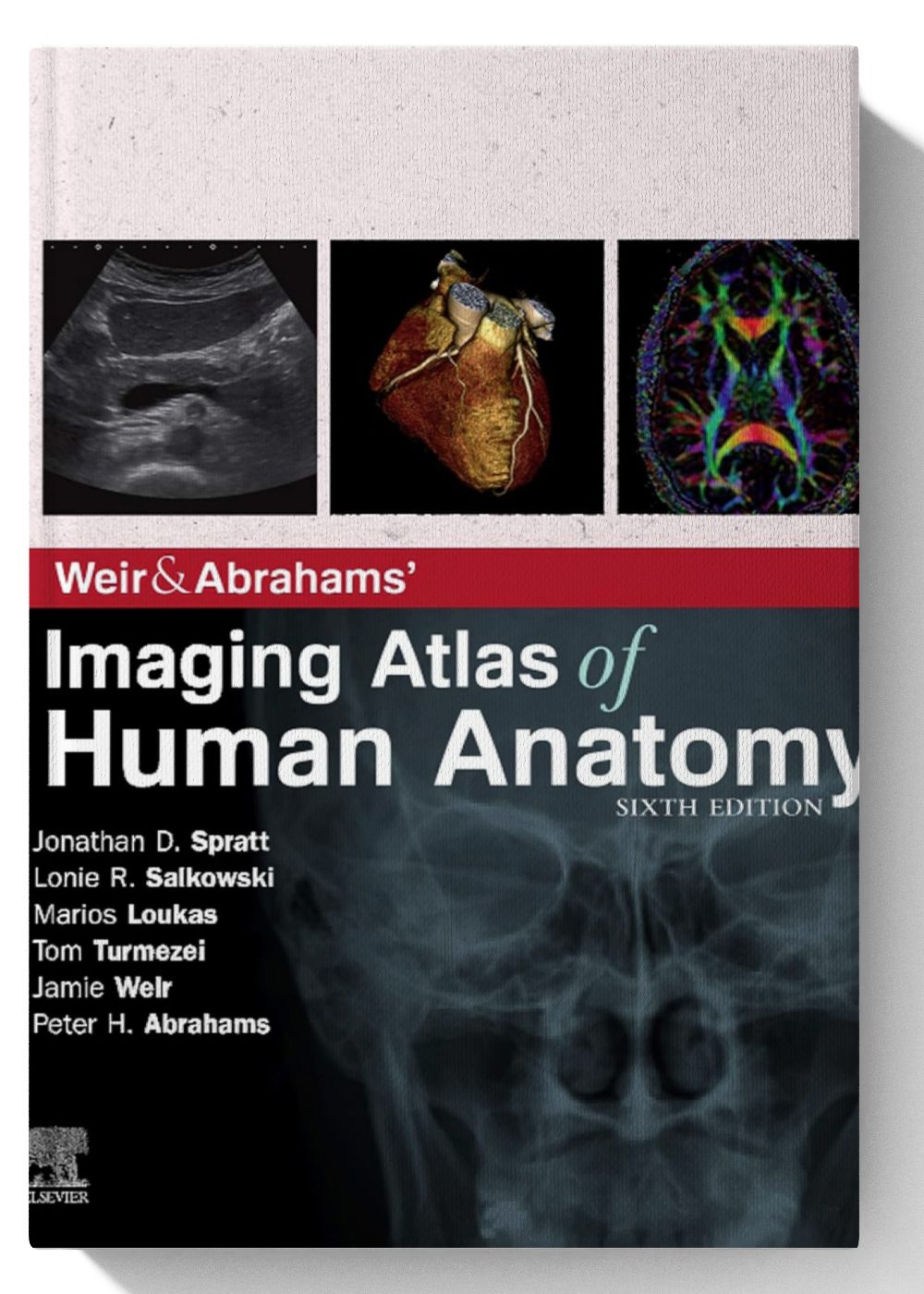
Weir & Abrahams’ Imaging Atlas of Human Anatomy E-Book, 6th Edition is a comprehensive and authoritative atlas of human anatomy using the latest imaging techniques. It is an essential resource for medical students, radiologists, radiographers, surgeons in training, and other healthcare professionals who need to understand the relationships between normal anatomy and the latest imaging modalities.
The book is divided into two parts: the first part covers general anatomy, including topics such as osteology, arthrology, myology, and neuroanatomy. The second part covers regional anatomy, including the head and neck, trunk, upper limbs, and lower limbs.
The book contains over 1,500 high-quality color images from a variety of imaging modalities, including plain radiographs, ultrasound, CT, MRI, and functional imaging. The images are labeled accurately and the text provides clear and concise descriptions of the anatomy.
Each chapter in the book begins with a learning objectives section, and ends with a key takeaways section. The book also includes a number of helpful features, such as clinical notes, bonus electronic content, and self-assessment questions.
Overall, Weir & Abrahams’ Imaging Atlas of Human Anatomy E-Book, 6th Edition is an excellent atlas of human anatomy using the latest imaging techniques. It is comprehensive, well-written, and well-organized. It is an essential resource for medical students, radiologists, radiographers, surgeons in training, and other healthcare professionals who need to understand the relationships between normal anatomy and the latest imaging modalities.
Here are some of the key features of the 6th edition:
- Over 1,500 high-quality color images from a variety of imaging modalities
- Learning objectives and key takeaways at the beginning and end of each chapter
- Clinical notes, bonus electronic content, and self-assessment questions
- Fully revised legends and labels, and new high-quality images
I hope this description is helpful. Please let me know if you have any other questions.


